UK Scientists Can Now Track Cancer Drugs Inside Living Cells For The First Time

Imagine knowing precisely where every cancer drug goes inside your cells. A groundbreaking new method could totally revolutionise cancer drug uptake understanding, by finally allowing scientists to see this vital process in living cells.
Game-Changing Discovery for Cancer Treatment
This new analytical method could improve how cancer treatments are designed. For the first time, scientists can track exactly where inside a living cell a drug accumulates. This breakthrough was made possible by researchers from King’s College London and the University of Surrey.
The technique detects trace amounts of metal inside individual living cells and their internal compartments. Crucially, it does this without needing to kill the cells first. The study, published in Spectrochimica Acta Part B, focused on targeted radionuclide therapy. This therapy works by attaching a radioactive particle to a molecule that seeks out tumour cells. It then delivers radiation directly to the cancer.
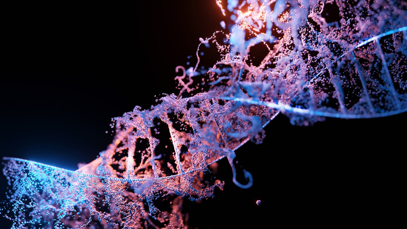
Where inside the cell the drug ends up is critical. A drug that reaches the nucleus causes damage to cancer by targeting DNA. Until now, there was no reliable way to measure this in living cells.
"We developed this method using two specialist facilities – the SEISMIC facility at King’s College London and the University of Surrey’s ICP-MS facility," explained Dr Monica Felipe-Sotelo, Senior Lecturer in Radiochemistry and Analytical Chemistry at the University of Surrey. "Together, they allowed us to combine the cell-sampling and metal-detection steps in a single workflow for the first time."
She added: "That combination is what makes it possible to ask not just whether a drug gets into a cell, but precisely where it goes once it’s there."
How It Works
The team used tiny glass capillary tips. These were ten micrometres wide for whole cells and three micrometres for subcellular structures. They extracted individual living pancreatic cancer cells and material from within them, including the powerhouse of cells, mitochondria, under a microscope.
King’s SEISMIC facility, funded by the Biotechnology and Biological Sciences Research Council, provided the specialist sampling capability. Surrey’s laser ablation inductively coupled plasma mass spectrometry (ICP-MS) facility then detected and measured thallium. This was done using LA-ICP-MS, a technique that uses a laser to vaporise minute quantities of material. A mass spectrometer then identifies and quantifies the metals within.
The combination of capillary sampling at the sub-cellular level and LA-ICP-MS had not been performed before. Researchers used thallium chloride as a chemically stable stand-in for thallium-201. This is a radioactive isotope currently under investigation as a cancer treatment candidate.
Thallium was successfully detected in individual cancer cells. It was also found, for the first time, inside mitochondria-enriched material extracted from those cells. This was achieved at extremely low amounts.
Thallium-201 is exciting as a potential cancer therapy because its radiation acts over such a short distance. This means it could destroy tumour cells while sparing the healthy tissue around them. But that precision cuts both ways: the drug has to end up in the right part of the cell to do its job.
Beyond Cancer: A Universe of Possibilities
The potential here goes well beyond cancer. Dr Claire Davison, Postdoctoral Research Associate at King's, and Dr Dany Beste, Senior Lecturer in Microbial Metabolism from the University of Surrey, highlighted its broad impact.
"The potential here goes well beyond cancer," they stated. "Metals play important roles in a wide range of diseases – from infectious disease to diabetes and liver conditions – and we have few tools for studying exactly where they are accumulating within cells."
They added: "This methodology gives us a way to do that with a level of precision and in conditions that are much closer to biological reality. That opens up a lot of questions we could not previously ask."
Professor Melanie Bailey, Professor in the Physical Sciences of Life at King’s, commented on the future. "We are continuing to develop this methodology at the SEISMIC facility and working with various different users to determine precisely where other drugs go when they enter cells, and what they do when they get there."
The technique could be extended beyond cancer research. It could study how any metal-based drug or toxic substance distributes inside living cells. Key next steps include extracting additional cellular compartments, like the nucleus. Improving methods to verify the purity of extracted subcellular material is also a priority.
Related Content
MORE: London scientists develop 'smart' drug that wipes out 95 per cent of cancer without toxic side effects — https://trendwiremedia.com/2026/03/07/london-scientists-develop-smart-drug-that-wipes-out-95-per-cent-of-cancer-without-toxic-side-effects/
MORE: Imperial College London and Oxford Create 'Digital Twins' to Fast-Track Life-Saving Drug Testing — https://trendwiremedia.com/2026/03/15/imperial-college-london-and-oxford-create-digital-twins-to-fast-track-life-saving-drug-testing/
MORE: New 'masked' cancer drug hides from healthy cells before killing tumours — https://trendwiremedia.com/2026/03/23/new-masked-cancer-drug-hides-from-healthy-cells-before-killing-tumours/
OFFICIAL SOURCE VERIFICATION: This report is based on official data from University Newsroom. Document: [New technique maps cancer drug uptake inside living cells | King's College London](https://www.kcl.ac.uk/news/new-technique-maps-cancer-drug-uptake-inside-living-cells) Source Link: https://www.kcl.ac.uk/news/new-technique-maps-cancer-drug-uptake-inside-living-cells
Author


Subscribe for $2 every four weeks for the first six months
Subscribe for $20 every four weeks for the first six months